Evolution of Under-Eye Care: From Home TV Routines to Modern Non-Invasive Tech

For decades, millions of viewers across Japan tuned in to NHK’s beloved infotainment flagship Tameshite Gatten for straightforward answers to stubborn physical woes. Among the show's most popular segments were everyday routines designed to tackle lower eyelid bags and persistent dark shadows, encouraging viewers to perform targeted facial muscle exercises to tone the orbicularis oculi. Viewers squinted into bathroom mirrors, hoping isometric contractions could reverse what felt like sudden aging. Today, the clinical conversation around periorbital aging has moved far beyond simple broadcast routines, as highlighted in a recent PR TIMES Report tracking peer-reviewed surgical techniques detailed in the plastic surgery journal PEPARS. Modern oculoplastic medicine is rewriting how clinicians and patients address under-eye hollows, trading television wellness hacks for targeted structural interventions.
📌 Key Takeaways:
- The Structural Limit: Facial muscle exercises popularized by broadcast programs build superficial muscle tone but cannot repair a stretched orbital septum or pull back herniated fat.
- The Anatomic Root: True under-eye bags stem from orbital fat prolapse combined with collagen depletion and tear trough ligament anchoring, creating harsh shadows that skincare creams cannot fix.
- The Modern Protocol: Patient demand has shifted toward non-invasive skin tightening like micro-focused ultrasound alongside sophisticated surgical structural shifts like the Hamra method.
The Broadcast Phenomenon That Defined At-Home Eye Care
During its multi-decade run, Tameshite Gatten earned immense viewer trust by testing health remedies through accessible science. When the program explored periorbital fatigue and swelling, it zeroed in on circulation and muscle tone. The logic sounded persuasive on television: training the ring-shaped orbicularis oculi muscle surrounding the socket should theoretically shore up sagging skin and clear pooled venous blood. Viewers across East Asia adopted these daily eye-narrowing drills as gospel.
The cultural appeal was obvious. Performing facial gymnastics cost nothing, carried zero surgical risk, and offered agency to people noticing their first signs of periorbital aging in their late thirties and forties. Cosmetic counters reinforced this mindset, selling firming eye creams alongside massage rollers designed to stimulate lymphatic drainage. For mild puffiness caused by poor sleep or fluid retention, these habits offered minor, temporary relief. Yet as years passed, millions of dedicated practitioners discovered their under-eye bulges remained stubbornly intact. The disconnect between at-home effort and biological reality prompted a deeper medical inquiry into why those bags formed in the first place.
Biological Realities of Orbital Fat Prolapse and Periorbital Aging
Clinical dermatology recognizes that under-eye bag causes are rooted in structural mechanics rather than weak surface muscles. The eye sits cushioned inside the bony orbit on a bed of three distinct intraorbital fat pads: medial, central, and lateral. Holding this fat safely inside the socket is a thin, fibrous membrane known as the orbital septum.
Periorbital aging involves several compounding breakdowns:
- Orbital Septum Laxity: Over time, the fibrous orbital septum loses tensile strength. Intraorbital fat pushes forward under gravity, herniating outward to create prominent bulges.
- Tear Trough Anchoring: Just below the herniated fat sits the orbicularis retaining ligament. This tough fibrous band tethers the skin tightly to the underlying facial bone, creating an unyielding groove known as the tear trough deformity.
- Dermal Thinning and Collagen Depletion: Lower eyelid skin measures under 0.5 millimeters thick, making it the most delicate skin on the human body. Natural collagen depletion strips this thin tissue of its remaining elasticity.
- Midface Volume Loss: As cheek fat pads descend with age, the structural foundation beneath the lower eyelid drops, widening the transition zone between the eye and midface.
When herniated fat casts a dark shadow into the tear trough groove below it, the eye looks perpetually exhausted. No amount of orbicularis oculi flexion can force herniated adipose tissue back behind a stretched fibrous wall. In fact, aggressive facial muscle exercises can repeatedly crease the thinning surface skin, accelerating mechanical skin laxity.
Evolving Protocols: Comparing At-Home Habits and Clinical Interventions
Navigating under-eye correction requires matching the intervention to the specific anatomical breakdown. Mild skin laxity responds differently than full-grade orbital fat prolapse.
| Treatment Approach | Primary Target | Duration of Results | Average Cost Range (USD) |
|---|---|---|---|
| Broadcast Muscle Drills & Creams | Superficial blood circulation and lymphatic drainage | Transient (a few hours) | $0, $120 |
| HIFU & RF Microneedling | Dermal collagen stimulation and shallow septum tightening | 6, 12 months | $600, $1,800 |
| Hyaluronic Acid Fillers | Camouflaging the tear trough groove below herniated fat | 9, 18 months | $700, $1,500 |
| Surgical Hamra Method (Blepharoplasty) | Transposing herniated fat pads to fill the orbital rim hollow | 10, 15+ years (permanent structural change) | $3,500, $7,500 |
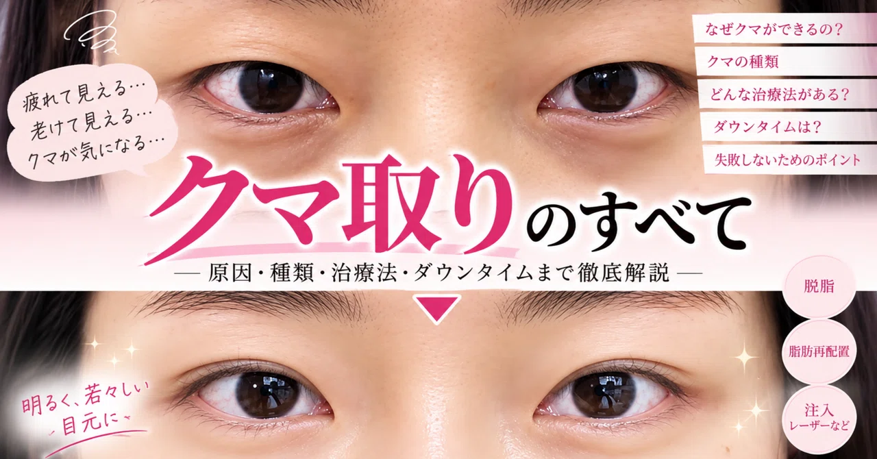
The Modern Surgical Standard: Fat Repositioning via the Hamra Method
Decades ago, conventional lower blepharoplasty relied on aggressive fat excision. Surgeons snipped away the bulging orbital fat pads through an incision below the lash line. While this flattened the bulge, it frequently left patients looking hollowed, skeletonized, and prematurely aged once the surrounding facial tissues continued to lose volume.
Surgical theory shifted dramatically with the widespread adoption of the Hamra method, originally pioneered by American plastic surgeon Dr. Norman Hamra and subsequently refined across specialty Asian practices. In his monograph for the plastic surgery journal PEPARS, Dr. Kazuhiko Maeda, Director of the Seishin Plastic Surgery Sapporo Clinic, highlighted how modernized approaches like the transcutaneous "Hamra plus alpha" technique solve the structural deficit. Rather than amputating the protruding fat, the surgeon releases the stubborn orbicularis retaining ligament and mobilizes the living fat downward over the lower orbital rim.
This maneuver accomplishes two mechanical goals simultaneously. It eliminates the protruding bulge while filling the sunken tear trough deformity with the patient's own vascularized tissue. By draping and securing this fat pedicle beneath the periosteum, surgeons create a flat, continuous contour from the eyelid down to the upper cheek. In patients over 50 who present with severe external skin laxity, modern transcutaneous variations also allow surgeons to carefully excise redundant crepey skin without pulling down the eyelid margin, preventing postoperative complications like ectropion.
The Non-Surgical Wave: Focused Ultrasound and Microneedling
Surgery delivers definitive structural realignment, but patient appetite for downtime has shrunk. For individuals in their late thirties and early forties displaying initial skin laxity rather than massive fat herniation, non-invasive skin tightening devices have become the treatment of choice.
Leading this clinical category are specialized high-intensity focused ultrasound (HIFU) handpieces engineered with narrow focal depths (1.5mm and 2.0mm) designed explicitly for periorbital anatomy. Devices utilizing linear ultrasound delivery rapidly heat targeted dermal zones to 65 degrees Celsius. This controlled thermal injury triggers an immediate contraction of collagen fibers followed by months of neocollagenesis, firming up the superficial layers beneath the eyes.
Complementing ultrasound is radiofrequency microneedling. By stamping insulated micro-needles directly into the dermis and releasing fractional radiofrequency energy, clinicians bypass the sensitive epidermal surface to stimulate deep structural remodeling. This approach addresses fine wrinkles and crepey dermal thinning that neither muscle exercises nor surgical fat repositioning can alter. Patients experience 48 hours of mild redness instead of two weeks of post-surgical bruising, making it an attractive maintenance protocol for early-stage periorbital aging.
Diagnosing the Patient: Ideal Candidates Versus High-Risk Profiles
The surge in clinic marketing requires careful patient screening. Dark under-eye circles fall into distinct diagnostic buckets, and misapplying treatments often worsens aesthetic outcomes.
- True Orbital Fat Prolapse (Ideal for Transconjunctival or Hamra Surgery): Evidenced by distinct bulges that become more prominent when looking upward. These patients gain zero long-term benefit from topical serums or muscle drills; surgical relocation of fat delivers an enduring structural fix.
- Structural Tear Troughs Without Fat Bags (Ideal for Fillers or Biostimulators): When hollows stem purely from genetic bone structure or fat loss, meticulously placed hyaluronic acid fillers or polynucleotide skin-boosters restore volume. However, injecting fillers in front of existing herniated fat is a critical mistake: it causes fluid retention, worsens heaviness, and creates the blue-tinted Tyndall effect.
- Dermal Laxity and Fine Crepiness (Ideal for HIFU and RF Microneedling): Patients whose primary complaint is loose, papery skin rather than protruding fat pads benefit most from energy-based collagen induction.
- Pigmentary or Vascular Dark Circles (Poor Candidates for Structural Surgery): If darkness stems from melanin deposits or hyper-visible microvasculature showing through ultra-thin skin, neither ultrasound nor surgery will erase the discoloration. These patients require chemical peels, vascular laser treatments, and targeted topicals like vitamin K and retinoids.
Frequently Asked Questions (FAQ)
Did the facial muscle exercises featured on broadcast TV shows actually work?
They helped stimulate microcirculation and temporary fluid drainage, which can briefly alleviate morning puffiness. However, they cannot structurally repair a stretched orbital septum or retract herniated orbital fat. In some cases, overworking the area accelerates fine dynamic lines in thinning skin.
What is the primary difference between standard fat removal and the Hamra method?
Standard lower blepharoplasty simply cuts out herniated fat, which risks creating a hollow, sunken eye socket over time. The Hamra method preserves the fat, releasing the tear trough ligament and shifting the fat downward to smoothly fill the sunken groove along the orbital rim.
Can non-invasive treatments like HIFU completely replace surgery?
No. Non-invasive devices excel at tightening loose dermal skin and stimulating collagen. They cannot move or shrink large volumes of herniated orbital fat. Once fat herniation becomes moderate to severe, surgical intervention remains the only anatomical solution.
The Road Ahead for Periorbital Longevity
The evolution of under-eye care marks a broader maturation in aesthetic medicine. The era of believing that a 10-minute television facial workout or a luxury peptide cream can reverse complex skeletal and ligamentous shifts has passed. Modern periorbital rejuvenation relies on diagnostic precision. Clinicians now evaluate the lower eyelid as a complex kinetic unit involving the orbital septum, dynamic retaining ligaments, localized fat pads, and fragile dermal layers. Whether a patient selects non-surgical collagen stimulation via focused ultrasound or structural fat repositioning through advanced Hamra protocols, lasting improvement comes from treating the exact anatomy causing the shadow.