Is Sleep Paralysis Dangerous? Fact-Checking Heart Strain, Suffocation, and Permanent Paralysis
Is Sleep Paralysis Dangerous? Fact-Checking Heart Strain, Suffocation, and Permanent Paralysis
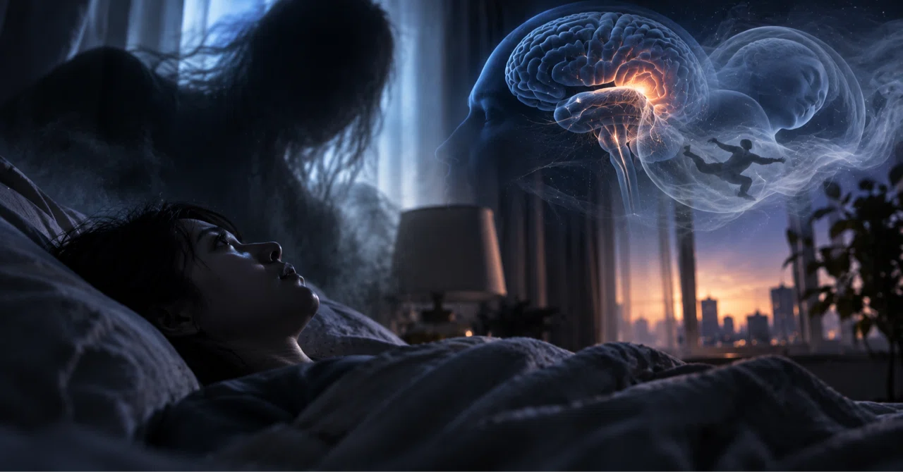
You wake up in the dead of night with your eyes open, your conscious mind fully alert, and your body locked in place. Your vocal cords refuse to fire, a crushing weight pins your ribcage, and a dark silhouette appears to linger just outside your peripheral vision. Across global cultures, this terrifying sensory trap has earned demonic folklore labels, most notably the Japanese phenomenon of kanashibari. As detailed in a clinical analysis by the medical commentary [note Report](https://note.com/noteigaku_hima/n/n47e0fee892c5), what feels like supernatural possession is actually a temporary misfire in brainstem signaling. Yet for millions experiencing it, the panic sparks an immediate biological question: can sleep paralysis stop your breathing, trigger a fatal heart attack, or leave you permanently paralyzed?
The short answer from contemporary sleep medicine is no. The sensation of imminent suffocation feels absolute, but the underlying mechanisms operate within standard mammalian neurology. Unpacking why this state feels lethal requires examining the boundary zone where rapid eye movement sleep overlaps with waking consciousness.
📌 Key Takeaways:
- Suffocation Myth: Autonomous diaphragm function remains intact through the phrenic nerve; chest pressure stems from relaxed intercostal muscles rather than airway collapse.
- Cardiovascular Impact: The adrenaline surge during an episode temporarily elevates heart rate and blood pressure, but healthy cardiovascular systems sustain no structural damage.
- Paralysis Permanence: Motor inhibition resolves entirely within seconds to several minutes as neurotransmitter release restores muscle tone.
- Primary Catalysts: Severe sleep deprivation, irregular shift schedules, supine sleep posture, and untreated obstructive sleep apnea represent the primary sleep paralysis causes.
The Biology of Kanashibari and REM Sleep Atonia
Sleep paralysis occurs when the brain wakes up before the body dismisses its nightly defense system. During rapid eye movement (REM) sleep, your brain dreams vividly while active motor neurons are shut down. This state, known as REM sleep atonia, prevents you from physically acting out dream scenarios and injuring yourself or your bed partner.
Two neurotransmitters, gamma-aminobutyric acid (GABA) and glycine, flood the motor neurons in the spinal cord, switching off skeletal muscle control. In normal sleep architecture, cortical arousal coincides precisely with the clearance of these neurotransmitters. During an episode of sleep paralysis, cortical awareness returns abruptly while the brainstem continues broadcasting the paralysis command. The result is waking up unable to move, completely conscious inside a non-responsive frame.
Because the brain remains partly anchored in REM neurochemistry, the visual and sensory centers keep generating dream imagery. These are known as hypnagogic hallucinations when falling asleep, or hypnopompic hallucinations when awakening. The brain senses total physical immobilization, interprets it as acute peril, and conjures an external predator or intruder to rationalize the threat.
Can Sleep Paralysis Stop Your Breathing or Trigger Suffocation?
The sensation of choking or having a heavy entity crushing the chest is the most distressing symptom of the condition. Patients frequently rush to emergency rooms convinced their respiratory system stalled mid-sleep. From an anatomical standpoint, complete asphyxiation during an episode is physiologically impossible in a healthy individual.
Human respiration relies on two separate muscular inputs: voluntary control and autonomic regulation. When you are awake, you actively use your intercostal muscles (the bands between your ribs) along with your diaphragm to modulate breath depth. During REM sleep atonia, voluntary skeletal muscles, including those intercostals, are temporarily shut down. Your primary breathing muscle, the dome-shaped diaphragm, is innervated by the phrenic nerve, which operates entirely outside the REM inhibition circuit.
Your diaphragm continues pulling air into the lungs uninterrupted. Because you cannot voluntarily take a deep, expanded chest breath, your conscious mind misinterprets shallow autonomic breathing as suffocation. Panic amplifies hypervigilance, converting normal shallow respiration into the tactile sensation of external chest pressure.
Cardiac Strain and the Myth of Sudden Heart Failure
When you discover you cannot move and perceive an intruder in the room, your amygdala triggers an immediate fight-or-flight emergency response. Epinephrine and norepinephrine pour into your bloodstream, sending your pulse soaring. For someone trapped in bed, a heart pounding at 130 beats per minute creates the visceral fear of an impending heart attack.
In individuals with a structurally sound heart, this transient sympathetic surge poses no more risk than a sudden jump scare during a horror movie or an intense sprint. The heart muscle handles acute sinus tachycardia routinely. The episode dissipates the moment motor tone snaps back into place, allowing parasympathetic pathways to reassert control and slow the heart down.
A legitimate clinical concern arises primarily in patients with pre-existing, advanced coronary artery disease, severe arrhythmias, or structural cardiac abnormalities. In those rare baseline conditions, extreme acute terror can theoretically induce ischemic stress. For the vast majority of the population, however, the episode leaves zero structural damage in cardiovascular tissue.
Isolated Episodes Versus Neurological Red Flags
Sleep medicine categorizes sleep paralysis into two distinct frameworks: isolated episodes and chronic symptoms tied to wider neurological sleep disorders. Occasional episodes require zero medical intervention, whereas frequent, debilitating recurrences demand specialized testing.
| Diagnostic Category | Typical Frequency | Primary Clinical Markers | Recommended Intervention |
|---|---|---|---|
| Isolated Sleep Paralysis (ISP) | 1, 3 times per lifetime | Triggered by acute exhaustion, alcohol intake, or jet lag; no daytime narcoleptic signs. | Reassurance, sleep debt recovery, sleep hygiene adjustments. |
| Recurrent Isolated Sleep Paralysis (RISP) | Multiple times per month | Persistent sleep-onset anxiety, severe insomnia, panic anticipations before bed. | Cognitive behavioral therapy for insomnia (CBT-I), low-dose REM-suppressing pharmacotherapy. |
| Narcolepsy Type 1 / Type 2 | Weekly or bi-weekly | Excessive daytime sleepiness, cataplexy (muscle collapse triggered by emotion), fragmented night rest. | Polysomnography, Multiple Sleep Latency Test (MSLT), orexin/hypocretin receptor modulators. |
| Sleep Apnea-Induced | Variable (1, 4 times monthly) | Loud snoring, waking gasping for air, high body mass index, micro-arousals during REM cycles. | Continuous Positive Airway Pressure (CPAP) therapy, oral appliance evaluation. |
Root Causes Driving Episode Frequency
The vast majority of episodes occur without any underlying neurological disease. Instead, they stem from physiological disruptions that fragment sleep cycles, increasing the odds that you wake up mid-REM.
Chronic sleep deprivation ranks as the single most common catalyst. When you consistently run on four or five hours of rest, your brain experiences REM rebound, an aggressive compensatory drive where REM cycles become deeper, longer, and more unstable. A sudden awakening out of rebound REM dramatically increases the odds of motor desynchronization.
Circadian rhythm disruption plays an equal role. Rotating night shifts, frequent cross-timezone travel, and wildly erratic sleep schedules scramble the brain’s internal suprachiasmatic nucleus. The brain loses its unified clock, allowing conscious awareness and motor atonia to overlap.
Sleeping flat on your back (the supine position) is another potent mechanical trigger. Clinical observations indicate that sleeping supine multiplies the likelihood of an episode up to fivefold compared to side-sleeping. Supine positioning allows the soft palate to collapse backward, creating upper-airway resistance. This minor obstruction triggers a micro-awakening in the cortex while the rest of the brain remains submerged in REM atonia.
Clinical Interventions and Daily Sleep Hygiene Protocols
Halting frequent episodes rarely requires intensive pharmaceutical intervention. In most clinical settings, correcting lifestyle habits dissolves the triggers that fracture sleep architecture.
Start by establishing a rigid circadian anchor. Go to bed and wake up at the exact same hour seven days a week, keeping bedtime variations within a 30-minute window. This consistency stabilizes REM timing and eliminates the erratic rebound phases that cause cortical disconnects.
Position training offers immediate relief for positional vulnerability. Sewing a small object into the back of a sleep shirt or using wedge pillows forces side-sleeping, keeping upper airways patent and lowering spontaneous REM micro-arousals. Eliminating alcohol within four hours of sleep prevents late-night REM fragmentation caused by ethanol rebound.
If you find yourself locked in an active episode, abandon the urge to fight the paralysis head-on. Straining large muscle groups, like trying to sit up or throw your arms, amplifies panic signaling in the amygdala, making the paralysis feel longer. Instead, focus entirely on the extremities exempt from heavy motor shutdown: blink your eyes rapidly, move your eyeballs side to side, or deliberately wiggle a single toe. These micro-movements send afferent sensory feedback back to the brainstem, breaking REM atonia within seconds.
Frequently Asked Questions (FAQ)
Q1: Can sleep paralysis cause permanent physical paralysis or brain damage?
No. The chemical blockade of motor neurons mediated by GABA and glycine is entirely reversible. The paralysis resolves on its own within a few seconds to three minutes, leaving zero motor impairment, structural nerve damage, or neurological deficit.
Q2: Why do episodes almost always involve shadow figures or an evil presence?
When the brain discovers it cannot move its body, it registers severe danger. The hyperactive amygdala searches for a threat to justify the paralysis. Combined with the dream projection centers active during REM sleep, your mind creates visual and auditory hallucinations, often taking the form of shadow figures, humming noises, or hovering entities.
Q3: When should someone see a sleep specialist for sleep paralysis?
Consult a physician or certified somnologist if episodes occur multiple times per month, cause persistent dread around going to bed, or are accompanied by overwhelming daytime sleepiness and sudden muscle weakness during laughter or surprise. Those overlapping signs warrant a formal evaluation for narcolepsy or sleep apnea.
What Lies Ahead for Sleep Paralysis Research
Neurologists now view sleep paralysis not as an esoteric medical anomaly, but as a window into the micro-circuits governing human consciousness. High-resolution functional neuroimaging continues mapping how thalamic switches desynchronize, demonstrating that wakefulness and sleep are not binary toggles, but overlapping neurochemical gradients.
For anyone waking up frozen beneath the phantom weight of an episode, remembering the biological facts is the fastest way to neutralize the fear. The chest pressure is an illusion of shallow autonomic breathing, your heart is beating against nothing more dangerous than an adrenaline surge, and your motor control will return within seconds. Treating the phenomenon as an internal timing error rather than a physical danger strips the night of its terror.
