Facial Expressions vs. Facial Structure: What Clinical Studies on Autism Actually Show

Short-form video feeds across TikTok, X, and Instagram are filled with creators claiming they can identify neurodivergence from a single selfie. Viral clips dissect eye tilt, jaw alignment, and resting expressions under search queries like "autism face" and "Asperger features." The rise of unauthorized third-party smartphone filters promising to screen users for developmental differences using facial scans has amplified this trend. These social media claims distort genuine clinical science into modern digital physiognomy.
Medical professionals are pushing back against the idea that autism spectrum disorder carries an obvious physical look. As detailed in a clinical review from a メディカルドック on MSN Report supervised by Dr. Ito, autism spectrum disorder and Asperger syndrome are diagnosed through behavioral, communication, and social interaction profiles, not visible facial markers. Conflating laboratory craniofacial research with everyday diagnostic reality creates anxiety for families while oversimplifying a deeply heterogeneous condition.
📌 Key Takeaways:
- The Diagnostic Reality: Autism spectrum disorder cannot be diagnosed from a photograph, mirror check, or consumer camera app.
- The Science: Specialized 3D craniofacial research detects microscopic phenotypic variations across small subsets of autistic children, but these millimeter-scale traits are invisible to the naked eye.
- Dynamic Differences: What observers mistake for an "autistic face" usually reflects dynamic differences in social reciprocity, spontaneous affect, or divergent eye gaze patterns.
- Clinical Harm: Relying on automated facial screening creates high rates of false positives and delays formal neurodevelopmental assessment.
The Social Media Pipeline Behind Viral Face Diagnostics
Online interest in physical markers of neurodivergence surged between 2024 and 2026 as algorithmic feeds rewarded quick diagnostic checklists. Online creators post side-by-side images comparing facial symmetry, midface depth, or perceived "flat expressions," telling viewers that these physical traits indicate undiagnosed Asperger syndrome or attention-deficit conditions. Comment sections quickly fill with anxious users evaluating their own childhood pictures.
The trend gained commercial momentum as independent developers launched amateur AI tools that claim to scan user facial geometry for developmental markers. These applications harvest user photos under the guise of accessible developmental screening. In reality, they evaluate static two-dimensional geometry using unvalidated training sets. Clinicians warn that these systems reproduce the pseudoscience of nineteenth-century phrenology, repackaged as modern machine learning.
Clinical assessments do not rely on still photographs. Board-certified clinicians evaluate social interaction over multiple hours through structured instruments such as the Autism Diagnostic Observation Schedule (ADOS-2). Reducing neurological variation to facial aesthetics strips away the behavioral context essential for clinical evaluation.
What Craniofacial Research Actually Measures
Viral posts often misquote legitimate developmental biology. The human face and the brain develop simultaneously during early embryonic gestation, sharing origins in the neural crest. Because of this shared developmental timing, genetic and developmental researchers study craniofacial morphology to identify early disruptions in brain development.
Landmark studies utilizing high-resolution stereophotogrammetry, advanced 3D surface imaging, have identified distinct facial morphology subgroups among autistic children. Researchers map thousands of facial coordinate points, measuring fractions of millimeters across the upper face, orbits, and philtrum. Findings reveal that small subgroups show subtle variations: a slightly wider upper face, shortened midface height, or minor changes in intercanthal distance.
These variations are not universal markers. They represent subtle phenotypic variations found across specific genetic subsets, such as children with co-occurring macrocephaly or rare monogenic syndromes like Fragile X or PTEN mutations. The naked eye cannot detect these sub-millimeter measurements on a playground, and commercial computer vision models cannot reliably isolate them from standard mobile phone selfies.
Dysmorphic Features Versus General Phenotypes
When developmental pediatricians conduct a clinical evaluation, they assess minor physical anomalies and dysmorphic features. These physical traits include low-set ears, epicanthal folds, unusual head circumference growth curves, or atypical hair whorls. These signs prompt genetic testing rather than a direct behavioral diagnosis.
Minor physical anomalies appear across many conditions, including isolated genetic anomalies and neurodevelopmental conditions, and they occur in neurotypical populations as well. The presence of a dysmorphic feature signals a potential systemic genetic variation; it does not confirm autism spectrum disorder. The diagnostic criteria defined in the DSM-5-TR and ICD-11 focus strictly on persistent challenges in social communication alongside restricted, repetitive patterns of behavior.
Most individuals diagnosed with autism spectrum disorder, including those historically diagnosed with Asperger syndrome, have completely typical facial structures. Confusing subtle physical anomalies with diagnostic traits leads parents down misleading rabbit holes while distracting from evidence-based behavioral therapies.
Dynamic Expression, Flat Affect, and Social Reciprocity
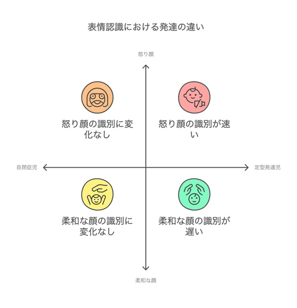
When laypeople insist they can "spot" autism in an individual, they are usually reacting to dynamic facial expressions rather than bone structure. Human interaction relies on rapid, nonverbal exchanges: fleeting smiles, eyebrow shifts, and mutual gaze adjustments. Autistic individuals often process and project these signals differently.
Autistic communication frequently involves divergent eye gaze patterns and variable facial expressivity. Some autistic individuals exhibit what observers describe as a flat affect, where spontaneous emotional states do not immediately translate into conventional facial movements. Others show intense, non-stereotypical expressions, or use expressive masking, consciously forcing conventional social expressions to blend into neurotypical environments.
These movements involve neurological signaling and social reciprocity, not skeletal structure. An autistic person resting their facial muscles appears distinct not because their facial bones differ, but because their dynamic interaction with social expectations runs on a different track. A static photograph cannot capture this complex neurological interplay.
| Diagnostic Dimension | Viral Claims & AI Scanners | Clinical Evidence & Research Reality |
|---|---|---|
| Facial Morphology | Claims unique, visible jawlines, eye shapes, or skull structures point to autism. | Identifies millimeter-level differences only through 3D photogrammetry in small genetic subsets; invisible to normal vision. |
| Facial Expressions | Interprets neutral photographs as proof of developmental differences. | Evaluates real-time social reciprocity, communicative timing, and dynamic affect over hours of observation. |
| Screening Technology | Uses unvalidated smartphone AI models that analyze 2D user portraits. | Applies validated behavioral screening (such as the M-CHAT-R) alongside developmental history and genetic assays. |
| Diagnostic Weight | Claims high standalone accuracy based on automated visual scans. | Carries 0% official diagnostic weight in DSM-5-TR; physical traits never confirm autism without behavioral data. |
The Clinical Risks of Photo-Based Self-Diagnosis
The drive to identify autism through photographs creates serious clinical issues. False positives induce unnecessary distress for families, sending parents on exhausting diagnostic journeys. Worse, false negatives can delay necessary care: a parent who assumes their toddler cannot be autistic because they lack a "supposed look" may ignore clear communication delays or motor challenges during key intervention windows.
Relying on physical traits also exacerbates racial and ethnic disparities in developmental healthcare. Facial morphology varies widely across global ancestries. Machine learning models trained on non-representative image databases regularly flag normal ethnic variations as dysmorphic features. Conversely, these algorithms often miss subtle developmental concerns in underrepresented groups.
Clinical evaluation requires holistic engagement. Developmental pediatricians, child psychologists, and speech-language pathologists look at communication milestones, sensory processing, and adaptive living skills. Bypassing this multidisciplinary process in favor of visual checklists delays targeted therapies that meaningfully improve life outcomes.
Frequently Asked Questions (FAQ)
Can an AI tool or mobile application diagnose autism from my photo?
No. Validated clinical protocols do not recognize standalone photo analysis as an autism diagnostic tool. Reliable diagnosis requires comprehensive neurodevelopmental assessment by qualified professionals who evaluate behavioral history, communication, and social interactions over time.
Why do some people say individuals with Asperger syndrome look alike?
Observers are often noticing dynamic behavioral cues rather than physical traits. Variations in eye contact, deliberate or unmasked facial expressions, and differences in conversational pacing can be misinterpreted as shared physical features, even though underlying craniofacial structures are completely typical.
What should I do if I suspect my child shows signs of autism?
Bypass social media checklists and speak with your pediatrician. Request a formal developmental screening using validated tools like the Modified Checklist for Autism in Toddlers (M-CHAT-R), followed by a comprehensive evaluation with a neurodevelopmental specialist.
Clinical Realities and Patient Care
The persistent belief that developmental differences write themselves across the face reveals our discomfort with invisible disabilities. In a visual culture shaped by social algorithms, people instinctively search for physical signs to categorize complex neurological traits. Autism spectrum disorder resists these simplistic visual shortcuts.
Autism encompasses diverse sensory profiles, communication styles, and cognitive frameworks. Research into craniofacial genetics offers valuable insights into early embryonic development, but it will never replace comprehensive behavioral evaluations. Prioritizing validated clinical evaluations over viral diagnostic shortcuts ensures that autistic individuals receive accurate assessments, meaningful support, and respectful care.