What Actually Happens Inside Your Spine When It Cracks: MRI and X-Ray Evidence
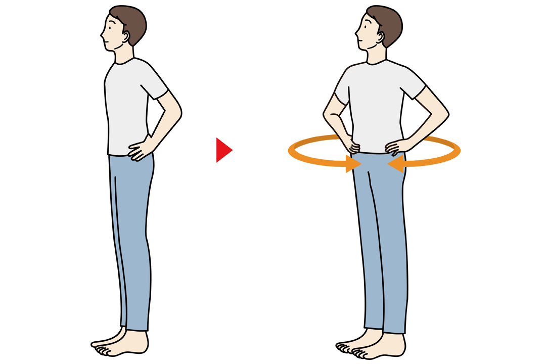
Every morning, millions of desk workers and gym-goers sit in an office chair, twist their torso sharply to one side, and wait for that resonant snap. The sensation produces an immediate, almost addictive wash of lower back stiffness relief. For decades, traditional medical dogma claimed that the cracking sound was simply the popping of microscopic bubbles inside the joint fluid. Recent diagnostic imaging reveals that the actual physics occurring within your lumbar spine is far more intricate, and far more demanding of caution.
Advances in high-speed, real-time magnetic resonance imaging (cine-MRI) have allowed biomechanical researchers to film spinal joint cavitation at dozens of frames per second. The resulting visual data overturns decades-old misconceptions about bone friction, disc alignment, and pressure drops inside the spinal column.
📌 Key Takeaways:
- The Core Mechanism: The auditory crack is not bone grinding against bone or a bubble bursting; it is the sudden formation of a low-pressure gas cavity within synovial fluid, a physical process known as tribonucleation.
- The Relief Illusion: The brief sense of decompression is largely neurological, caused by a temporary reset of the surrounding paraspinal muscle spasm rather than permanent vertebral realignment.
- The Long-Term Hazard: Habitual, unguided self-cracking frequently targets already hypermobile segments, accelerating ligamentous joint laxity while leaving stiff, restricted joints immobilized.
Inside the Synovial Void: Real-Time Imaging of Cavitation
The human lower back contains five lumbar vertebrae connected posteriorly by pairs of zygapophysial joints, commonly called facet joints. Each facet joint is encased in a fibrous capsule filled with thick, lubricating synovial fluid. When you apply rotational force or lateral flexion to your torso, you pull those joint surfaces apart.
In a landmark imaging protocol led by Dr. Gregory Kawchuk at the University of Alberta, researchers rigged a cable system inside an MRI suite to pull fingers and joints into cavitation while continuous cine-MRI recorded the event. The scans settled an eighty-year medical debate. As the opposing articular surfaces separate under tensile load, the adhesive resistance of the synovial fluid fights the pull until a critical threshold is reached.
At that exact millisecond, the internal hydrostatic pressure drops catastrophically. The sudden negative pressure causes dissolved gases, predominantly carbon dioxide and nitrogen, to come out of solution instantaneously. This process, known as tribonucleation, produces a rapid creation of a gas-filled cavity inside the joint capsule. That rapid vacuum formation generates the sharp acoustic crack.
The cavity does not vanish immediately. Post-cavitation imaging demonstrates that these synovial fluid gas bubbles linger inside the joint space for up to 20 to 30 minutes. During this window, known as the refractory period, the joint cannot be cracked again because the dissolved gas has not yet reabsorbed into the liquid medium.
Why Lumbar Decompression Triggers Immediate Muscle Relaxation
If the pop is just a micro-vacuum of nitrogen gas, why does it deliver such profound lower back stiffness relief? The answer lies in neurophysiology rather than skeletal architecture.
The fibrous capsule enclosing each facet joint is saturated with mechanoreceptors, specifically Type I and Type II nerve endings that monitor tension, position, and mechanical pressure. When the joint surfaces rapidly gap during cavitation, these receptors fire a massive volley of afferent signals into the posterior horn of the spinal cord. This input triggers a local inhibitory reflex that downregulates hyperactive motor unit firing in the surrounding multifidus and erector spinae muscles.
A sustained paraspinal muscle spasm is essentially short-circuited. The muscle fibers relax, local capillary blood flow increases, and the patient experiences a transient drop in pain levels via the gate control mechanism of nociception. You feel lighter and more mobile not because a "slipped bone popped back into place," but because your central nervous system temporarily turned down the muscular armor guarding the joint.
Biomechanical Comparison: Self-Manipulation vs Clinical Intervention
Spinal movement exists on a spectrum from active mobilization to high-velocity force. Understanding the structural differences between uncontrolled self-cracking and targeted clinical therapy explains why chronic self-manipulators often worsen their own symptoms over time.
| Intervention Method | Biomechanical Target | Cavitation Frequency | Tissue Strain & Instability Risk |
|---|---|---|---|
| Seated Torso Twisting | Broad rotational torque across L1, L5; path of least resistance | 60%, 85% per attempt | High: Induces torsional shear on annular disc fibers |
| Foam Rolling Lumbar Spine | Compressive hyperextension over lumbar lordosis | 25%, 40% per attempt | Moderate to High: Jams facet joints together under bodyweight |
| Targeted Chiropractic Spinal Manipulation | Isolated, segment-specific high-velocity low-amplitude thrust | 70%, 90% at targeted joint | Low to Moderate: Pre-positioned to lock out adjacent mobile segments |
| Axial Decompression & Mobilization | Passive longitudinal traction; non-rotational intervertebral gapping | 10%, 20% (cavitation is incidental) | Minimal: Preserves ligamentous tension without shear load |
The Hypermobility Trap and the Pathology of Chronic Cracking
The biomechanical danger of self-cracking lies in the law of least resistance. The human spine functions as a kinetic chain. When a patient develops lower back stiffness, that rigidity is almost never uniform. Typically, one or two segments, often the lower thoracic vertebrae or the L5, S1 junction, are hypomobile, stiffened by postural strain or microtrauma.
When you sit in a chair and wrench your shoulders around to force a pop, you cannot direct the torque into a frozen spinal segment. Instead, the force naturally channels into the most flexible, easily moveable joint immediately adjacent to it. You pop the segment that is already working too hard.
Doing this five to ten times a day triggers progressive joint laxity. The facet capsular ligaments, the ligamentum flavum, and the interspinous ligaments stretch out microscopically. Over months and years, hypermobility and joint laxity set in at the overstretched levels. Because these loose ligaments can no longer provide structural stabilization, the central nervous system compensates by locking down the paraspinal muscles even tighter.
The individual feels stiff, twists aggressively to crack the joint for relief, and temporarily calms the muscle spasm, only for the neurological tension to return hours later with increased intensity. It is an insidious physiological loop.
Rotational Shear, Annular Tears, and Disc Herniation Risks
The lumbar spine is anatomically built for flexion and extension, not extensive rotation. While thoracic facet joints sit in a coronal plane that permits rotational movement, lumbar facet joints face largely inward in a sagittal orientation. Their primary architectural job is to stop your lower back from twisting too far.
Forcing deep rotational pops introduces torsional shear stress across the intervertebral discs. The annulus fibrosus consists of roughly 15 to 25 concentric sheets of collagen fibers arranged in alternating diagonal angles. During twisting, only half of these fibrous layers are oriented to resist the load, effectively cutting the disc's mechanical strength in half.
Aggressive rotational self-manipulation generates acute lumbar disc herniation risks. If a micro-tear already exists within the posterior annular wall, high-torque twisting pushes the pressurized nucleus pulposus outward through the defect. This mechanical extrusion can quickly develop into nerve impingement symptoms, radiating burning pain, numbness, and motor deficits down the pathway of the sciatic nerve.
Self-manipulators also regularly mistake sacroiliac joint dysfunction for spinal tightness. The sacroiliac joint connects the sacrum to the pelvic bones with dense, rigid ligaments designed to transfer walking loads, not to pop freely. Forcing an SI joint to crack through pelvic contortions frequently destabilizes pelvic alignment, aggravating deep buttock ache that resists conventional treatment.
Safer Protocols for True Decompression and Mobility
Breaking the compulsive cracking habit requires replacing violent joint manipulation with active decompression and muscular balance. Achieving authentic lumbar spine decompression does not require a loud acoustic report.
Patients dealing with chronic tightness should prioritize pure axial unloading. Hanging from an overhead pull-up bar with feet lightly resting on the floor allows bodyweight to exert low-load, long-duration traction across the lumbar lordosis. This expands the intervertebral foramina and drops intradiscal pressure without applying dangerous rotational torque to the annular fibers.
Foam rolling requires equal caution. Placing a hard cylinder directly beneath the lower back and rolling aggressively forces the lumbar spine into excessive hyperextension, jamming the inferior and superior articular facets into each other under bodyweight. Instead, therapists recommend positioning the foam roller beneath the mid-to-upper thoracic spine or across the gluteal complex and hip flexors. Freeing restriction in the hips and mid-back removes the compensatory mechanical strain that makes your lower spine feel compressed in the first place.
Frequently Asked Questions (FAQ)
Q1: Does cracking your lower back cause arthritis?
A1: Long-term observational studies on knuckle cracking showed no direct causal link to osteoarthritis, but the spine bears heavy axial weight that the hands never encounter. Chronic, forceful spinal cracking that induces ligamentous laxity can lead to compensatory bony remodeling, facet joint hypertrophy, and accelerated degenerative wear over several years.
Q2: Why does foam rolling my lower back feel unstable or painful?
A2: The lumbar spine has no protective rib cage to limit excessive backward bending. When you roll directly across your lumbar region, the roller drives the spine into hyperextension, crushing the facet joint capsules and straining the anterior longitudinal ligament. Focus rolling on the thoracic spine and glutes instead.
Q3: How do I know if my back pain is coming from a disc rather than a tight joint?
A3: Pure joint stiffness generally stays localized in the lower back and improves with gentle, active walking. A developing disc issue frequently manifests as a sharp, central ache that worsens during sitting, bending forward, coughing, or sneezing, and may radiate pain, tingling, or weakness into the buttocks, thighs, or calves.
Q4: How long does a facet joint take to reset after popping?
A4: The refractory period typically lasts between 15 and 30 minutes. During this window, the released gas bubbles remain in the joint capsule, making another cavitation mechanically impossible until the gas dissolves back into the synovial fluid.
Navigating Spinal Stability in Modern Movement Practice
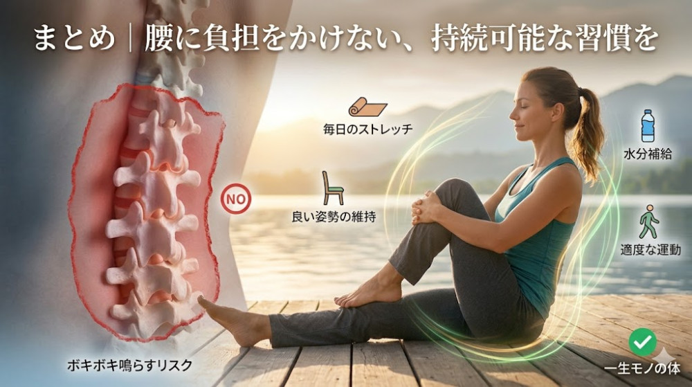
The auditory pop of a facet joint release offers short-term psychological satisfaction and transient neurological relief, but it is not a cure for chronic postural overload. Equating joint cavitation with structural health ignores the delicate biomechanical balance between mobility and stability.
When the lumbar spine feels persistently jammed, the real solution rarely involves forcing another sharp twist. Sustained functional relief comes from stabilizing hypermobile segments with isometric core bracing, opening stiff hip joints, and decompressing the intervertebral spaces through gentle axial traction. Your lower back does not need louder cracks; it needs an environment where its ligaments can finally hold their ground.